Alpha Theta Delta and Beta: Descriptions Not Buttons
Alpha, beta, theta and delta are useful EEG labels—not buttons for focus, creativity or sleep. Learn why boundaries vary, rhythms coexist and context changes what a band can mean.
 Jonas ReedCalm Signal editor5 September 2026
Calm Signal · Visual guide
Alpha, beta, theta and delta are useful labels for patterns in an EEG. They are not buttons that switch the brain into creativity, sleep or perfect concentration.
In short
- Frequency bands describe how quickly measured activity oscillates; several bands are present at the same time.
- Band boundaries are conventions and differ between laboratories, devices and individuals.
- A band does not have one fixed psychological meaning. Alpha, for example, changes with eyes, attention, location and task.
- Consumer audio or wearables cannot guarantee a desired state simply by displaying or playing a frequency.
Start with the signal, not the label
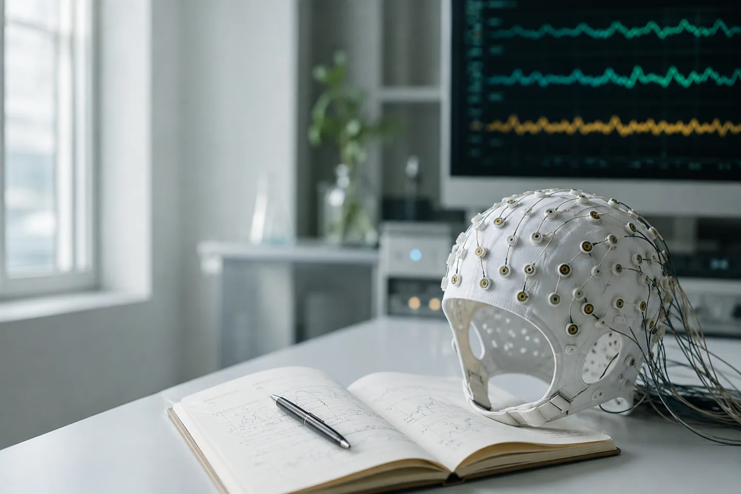
Electroencephalography records tiny changes in electrical potential from electrodes placed on the scalp. Those changes mainly reflect coordinated activity across populations of cortical neurons. EEG is excellent at showing when activity changes, but the skull and surrounding tissues blur where it originated.
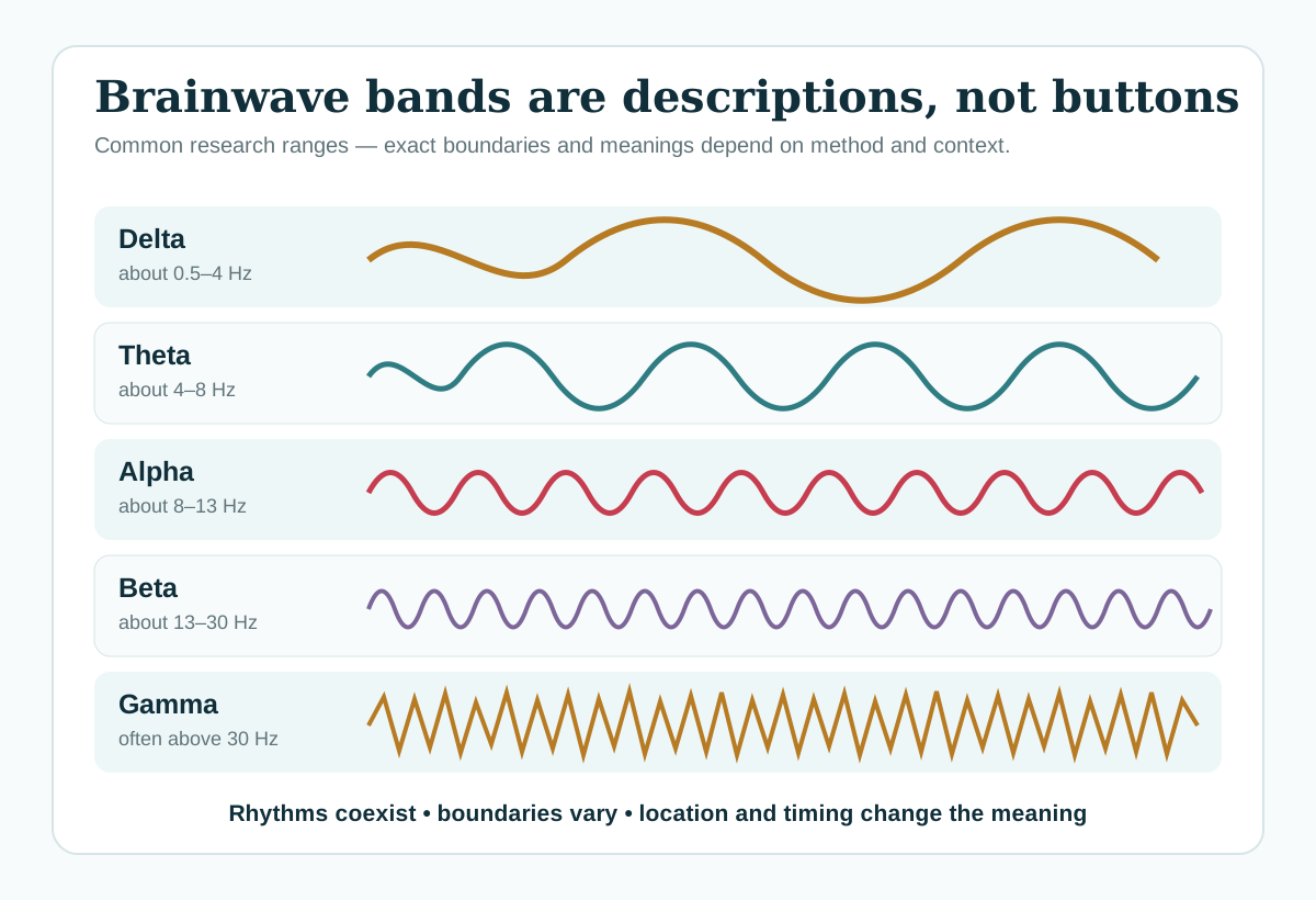
Researchers can describe a signal in the time domain—how voltage changes moment by moment—or in the frequency domain—how much activity occurs at different repetition rates. One hertz means one cycle per second. Dividing the spectrum into named bands makes results easier to compare, but nature does not place a border at exactly 8 or 13 Hz.
What the familiar bands can describe
| Band | Common approximate range | Contexts in which it is studied |
|---|---|---|
| Delta | 0.5–4 Hz | Deep NREM sleep, slow cortical activity and some clinical patterns |
| Theta | 4–8 Hz | Drowsiness, memory, navigation and cognitive control |
| Alpha | 8–13 Hz | Eyes-closed wakefulness, sensory inhibition and attention |
| Beta | 13–30 Hz | Active processing, movement and sensorimotor networks |
| Gamma | Often above 30 Hz | Local processing, attention and integration across neural populations |
These associations are starting points, not translations. Theta during a memory task is not automatically the same phenomenon as theta near sleep. Beta over sensorimotor cortex is not a generic “busy mind” meter. Gamma recorded at the scalp can be difficult to separate from small eye, forehead, jaw and neck muscle signals.
Alpha is the clearest example of context
Close your eyes while awake and relaxed and activity near the alpha range often becomes more prominent over posterior scalp regions. Open your eyes or direct visual attention and that activity may decrease. Yet alpha can increase in one brain region while decreasing in another, and its peak frequency differs across people and across the lifespan.
A classic review of alpha and theta research argued that fixed bands can hide important differences and that an individual’s alpha peak may provide a better reference. More recent methodological guidance adds another complication: part of the measured spectrum is non-oscillatory background activity. A rise in “band power” can therefore occur without a tidy, sustained rhythm appearing.
Brain rhythms are usually bursts and mixtures
Simple diagrams show smooth sine waves because they teach frequency clearly. Real neural oscillations can be brief, irregular and non-sinusoidal. Their shape can produce harmonics—extra frequency components that look like separate rhythms. Two sources can also overlap at the same electrode.
Good analysis asks whether an oscillation is truly present, whether the chosen band fits the individual data, how long the activity lasts, where it occurs and whether movement or muscle artefact could explain it. The coloured bar on a consumer dashboard usually hides those decisions.
Why “increase alpha” is not a complete goal
A claim may move from “alpha changed during a task” to “alpha produces relaxation” and then to “this product produces alpha, therefore it relaxes you.” Each step requires new evidence. A sound pulsing ten times per second is an acoustic modulation; it is not itself an alpha brainwave. Even if an EEG response occurs at that rate, the response does not establish a meaningful benefit.
The same problem applies to beta for focus, theta for creativity or delta for healing. A useful outcome should be measured directly: did attention improve on a validated task, did sleep improve over time, or did distress decrease compared with an appropriate control?
How to read a band-based claim
- Ask where the signal was measured. Scalp location and sensor type matter.
- Check the comparison. Eyes open versus closed, rest versus task and wake versus drowsiness are not interchangeable.
- Look for individual variation. A fixed range may miss a person’s actual peak.
- Separate entrainment from outcome. A frequency response is not proof of better mood, sleep or performance.
- Check artefact handling. High-frequency findings especially need convincing muscle and movement controls.
Sources and further reading
- Donoghue et al. (2022): Methodological considerations for studying neural oscillations
- Klimesch (1999): EEG alpha and theta oscillations in cognition and memory
- Muthukumaraswamy (2013): High-frequency MEG/EEG activity and muscle artefacts
- American Academy of Sleep Medicine: Sleep-stage scoring update summary