Sleep Spindles: What They Reveal About Memory and Rest
Sleep spindles are brief EEG bursts linked with thalamocortical coordination. Memory research points to their timing with slower and faster rhythms—not simply a higher count.
 Elena MoralesSleep & Recovery editor5 September 2026
Sleep & Recovery · Sleep glossary
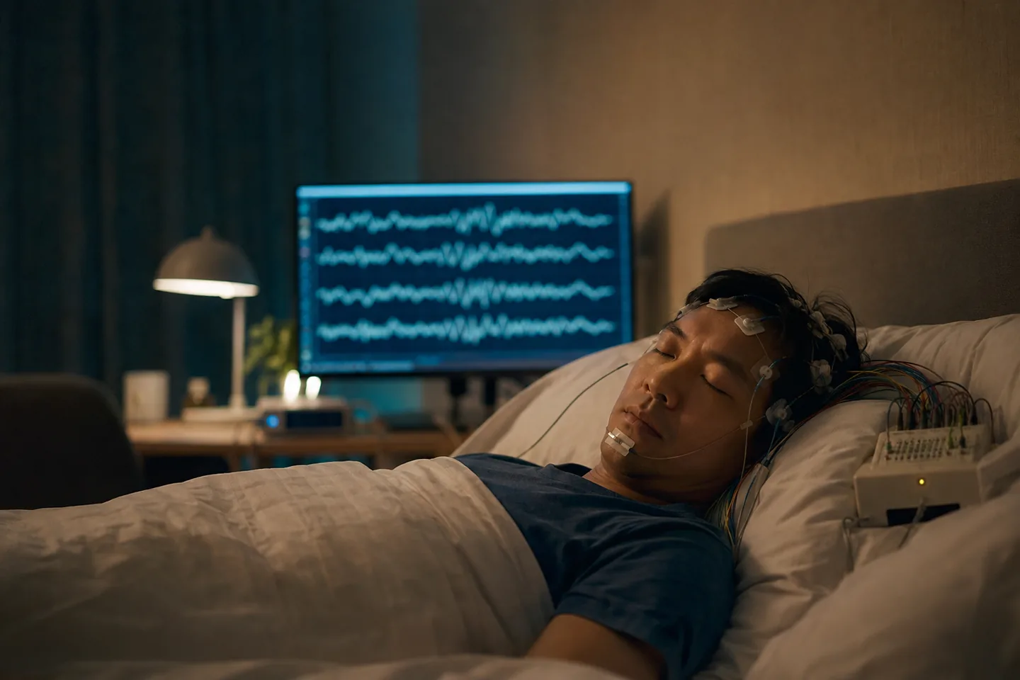
Sleep spindles are brief bursts of rhythmic EEG activity, usually seen during N2 sleep. They reflect thalamocortical coordination and are studied in sleep stability, learning and memory—but a higher nightly count is not a universal target.
In short
- A spindle is a waxing-and-waning burst, commonly around 11–16 Hz, lasting roughly half a second to two seconds.
- Spindles arise through interactions between the thalamus and cortex and help define N2 sleep.
- Memory research focuses on when spindles occur relative to slow oscillations and hippocampal ripples, not simply how many appear.
- Consumer wearables do not directly measure spindles unless they include suitable EEG sensors and validated analysis.
What a spindle looks like
On the scalp EEG, a sleep spindle looks like a compact packet of waves that grows and then fades—hence the name. The American Academy of Sleep Medicine uses activity around 11–16 Hz, lasting at least 0.5 seconds, as a characteristic feature of N2 sleep. Researchers also discuss slower frontal and faster central or parietal spindle activity, although definitions and detection algorithms vary.
A spindle is not a miniature period of waking and is not normally something a sleeper can feel. It is a surface sign of coordinated activity in thalamocortical circuits: loops connecting the thalamus, which relays and regulates information, with the cerebral cortex.
How the thalamus helps generate the burst
During non-REM sleep, thalamic reticular and thalamocortical neurons can enter a rhythmic mode. Their reciprocal inhibition and rebound firing create oscillatory bursts that reach the cortex. Spindles can be local or widespread, and their expression changes across the night, brain regions, age and individuals.
They are also among the more heritable features of a person’s sleep EEG. That stability is one reason cross-sectional correlations with cognitive ability must be interpreted carefully: a spindle measure may reflect anatomy, development and stable individual differences as well as what happened that day.
Sleep stability and sensory gating
The thalamus reduces the flow of some sensory information to cortex during sleep, and spindles have been linked with periods of reduced responsiveness. This may help ongoing sleep resist irrelevant input. Yet the sleeping brain is not sealed off; meaningful or intense stimuli can still be processed and can trigger arousal.
It is more accurate to describe selective gating than a protective wall. A quiet, safe bedroom supports sleep whether or not a device claims to increase spindles.
Why spindles appear in memory research
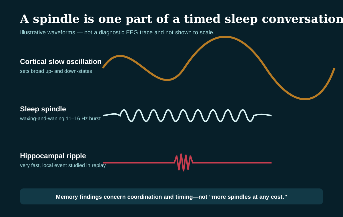
One influential model proposes that recent memories are reactivated during non-REM sleep. Cortical slow oscillations provide broad timing, hippocampal sharp-wave ripples carry brief replay-related activity, and thalamocortical spindles help coordinate communication and plasticity across regions.
A recent Bayesian meta-analysis of 23 studies found strong evidence that precise, strong coupling between slow oscillations and fast spindles in frontal regions predicts memory retention. The association varied with memory type, ageing, frequency and topography. This supports a timing mechanism; it does not mean that adding undifferentiated spindle activity guarantees better memory.
Count, density and coupling answer different questions
| Measure | What it describes | Main caution |
|---|---|---|
| Count | Number of detected events | Depends on sleep time and detection threshold |
| Density | Events per minute of a stage | Algorithms and electrode locations differ |
| Amplitude or power | Size of spindle-range activity | Skull, sensors and background spectrum affect it |
| Coupling | Timing relative to a slow oscillation or ripple | Analysis choices can change the estimate |
What ageing and illness can change
Spindle characteristics change across development and often decline or shift with ageing. Differences are also studied in neurological, psychiatric and sleep conditions. These are group-level research patterns, not standalone diagnoses. A clinician interprets EEG events alongside sleep stages, symptoms, medication, breathing and other signals.
Can sound increase spindles?
Laboratories have tested sounds timed to the phase of slow oscillations. Some experiments report changes in slow waves, spindles or memory, while results depend on timing, intensity and participant. Closed-loop stimulation detects the ongoing EEG and adapts moment by moment. A normal audio track does not know the sleeper’s phase and should not be described as doing the same thing.
The practical route to memory-supportive sleep remains adequate opportunity and treatment of sleep disorders. Loud snoring, breathing pauses, severe daytime sleepiness, persistent insomnia or unusual night behaviour merit clinical attention; a spindle score does not rule them in or out.
Sources and further reading
- Fernandez and Lüthi (2020): Sleep spindles—mechanisms and functions
- Ng et al. (2025): Slow-oscillation–spindle coupling and memory meta-analysis
- Antony et al. (2019): Sleep spindles and memory reprocessing
- Coenen (2024): Sensory gating and gaining in sleep
- American Academy of Sleep Medicine: Sleep-stage scoring update summary